
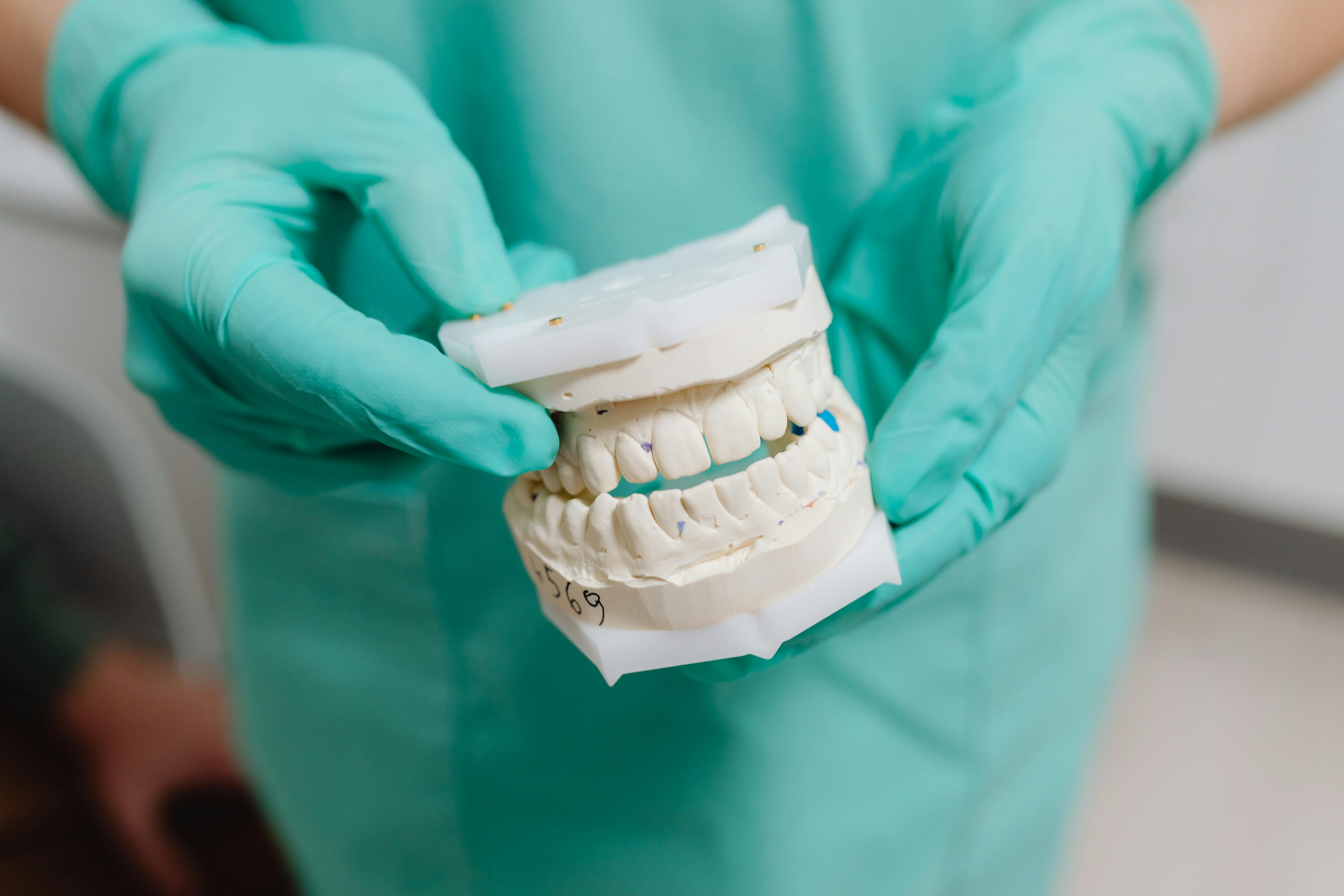
The color of a tooth is set largely by the layer underneath its surface rather than by the surface itself. That arrangement explains why some cosmetic requests are straightforward and others are not.
Enamel behaves like frosted glass
Enamel is a dense mineral shell that transmits and scatters light rather than blocking it. Light passes partway through, reflects off the deeper tissue, and travels back to the eye.
Because of that behavior, enamel works more like frosted glass than like paint. Its thickness and crystal structure change how much of the underlying color reaches an observer.
At the biting edges of front teeth, enamel is not backed by dentin, so light passes almost straight through. Those edges often read as grayish or faintly transparent as a result.
Dentin supplies most of the visible color
Dentin sits beneath enamel and is naturally warmer, running through cream and light brown tones. Its color is a property of the tissue itself, not a deposit sitting on the surface.
Dentin is also arranged in microscopic tubules that run outward from the pulp. That structure affects how light scatters, which is part of why teeth rarely look like a flat block of color.
Since the dominant color source lies below the surface, polishing or scrubbing the outside changes very little. It removes surface film without touching the layer doing most of the work.
Why teeth commonly look darker with age
Enamel thins gradually through ordinary chewing, acid exposure and brushing over decades. As the outer layer becomes thinner, more of the warmer dentin color is visible through it.
At the same time, the tooth continues laying down secondary dentin on the inside of the pulp chamber. The dentin layer effectively grows thicker while the enamel over it grows thinner.
Both changes push in the same direction, which is why older teeth tend to appear deeper in tone. It is a structural shift rather than accumulated dirt.
How cosmetic planning accounts for the layering
Restorative materials are built to imitate this layered optical behavior instead of matching a single flat shade. Ceramics and composites are made in body and enamel layers with different translucency.
A dentist assessing a front tooth is judging opacity and light transmission, not only hue. A restoration that matches on a shade tab can still look wrong once light passes through it.
The thickness available also constrains the result. A thin veneer over a dark tooth may not mask the underlying color, which changes what materials and preparations are workable.
Where a clinical assessment is the deciding step
Discoloration in a single tooth can reflect an internal change, such as trauma affecting the pulp, rather than surface staining. Those situations are diagnosed in an office, not by appearance alone.
An examination with radiographs establishes whether the tooth is sound before any cosmetic work is considered. That order matters because appearance work does not address underlying disease.
Anyone weighing a change in tooth color should raise it with a licensed dentist, who can identify what is producing the color in that particular mouth.